Overview of Services
The Comparative Medicine Animal Histology Service (CMAHS) Center provides a comprehensive range of histological research techniques that include:
- Standard tissue processing and paraffin embedding
- Standard and oversized specimens
- Special orientation of tissues
- Including orientation to match images from MRI, CT, and other modalities
- Special consultation required
- Serial sectioning, step sectioning, and sectioning of frozen specimens
In addition to routine hematoxylin and eosin staining (H&E) of tissue sections, we offer a variety of special stains to highlight specific cell types, cellular elements, tissue components, or infectious organisms. Additionally, we offer immunohistochemical / immunoperoxidase and apoptosis assays.
The Comparative Medicine Animal Histology Service Center is located on the third floor of the Edwards Building in room R330.
Details of services provided through the CMAHS Center
Tissue processing ONLY
- Tissue processing encompasses the preparation of previously fixed tissues for paraffin embedding.
- Tissue processing involves a series of dehydration, clearing, and paraffin (wax) embedding steps.
- We offer a "processing only" service, which allows investigators to embed the processed tissues themselves so they can spatially orient the specimens in a way that best suits their specific experimental needs.
Tissue processing AND embedding
- After the tissues are processed, they can be embedded in paraffin to make paraffin tissue blocks, that can then be sectioned with a microtome.
- We offer tissue processing and paraffin embedding services with the final result being a paraffin tissue block.
- Additionally, we offer a variety of sectioning and staining services including: 1) the production of unstained slides, 2) routine H&E staining, 3) histochemical ("special") stains, and 4) immunohistochemistry / immunoperoxidase. Please see the following sections for a brief explanation of these services.
H&E staining
- Hematoxylin and eosin (H&E) staining is one of the principal and most widely used stains in histology and is generally regarded as the "gold standard" for evaluation of bright field histology specimens.
- Additionally, evaluation of H&E preparations is necessary for providing a context in which to adequately interpret observations made in other specialized staining techniques such as histochemical ("special") and immunohistochemical stains.
- We provide H&E staining for the tissues and slides we process, or as a stand-alone service (H&E only) on unstained slides of either paraffin-embedded or frozen sections.
Re-cuts with or without staining
- Once paraffin tissue blocks are made (see "Tissue processing and embedding" above), they can be sectioned with a microtome multiple times for a variety of purposes.
- Some of the uses for re-cuts include: 1) production of unstained slides for staining at a later time, 2) routine H&E staining, and/or 3) other advanced staining techniques (histochemical "special" stains and/or immunohistochemistry).
- We offer microtome sectioning services ("re-cuts") of paraffin blocks with or without additional staining, as well as serial and step sectioning.
Histochemical ("special") stains
- Histochemical ("special") stains aid in the visualization and/or identification of structures and/or substances in tissue sections.
- Different special stains can be used to identify cellular components (e.g. intra-cellular lipid, intercalated discs in cardiac cells), cellular accumulations (e.g. iron, copper), matrical and connective tissue elements (e.g. collagen, elastin), and infectious elements (e.g. fungi, bacteria, protozoa).
- Please consult with us for the most appropriate stain(s) for your experimental needs.
- We strongly recommend always doing a paired H&E-stained section (gold standard) to provide a context in which to interpret observations made in histochemical preparations.
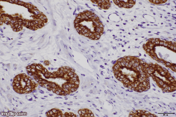
Immunohistochemistry
- Immunohistochemistry is based on the principle of antibody binding, and is used to identify specific antigens and specific antigenic determinants (epitopes) in tissue sections.
- Immunohistochemistry allows for identification and differentiation of specific cell types (e.g. smooth muscle cells, T-lymphocytes, endothelial cells), cellular components (e.g. mitochondria), and specific matrical and connective tissue elements (e.g. collagen IV).
- We offer antibody optimization and antibody staining services.
- We strongly recommend always doing a paired H&E-stained section (gold standard) to provide a context in which to interpret observations made in immunohistochemical preparations.
Getting Started
Publication Acknowledgement
As with all Stanford Service Centers, credit must be given to the Comparative Medicine Animal Histology Service Center for data that results in a publication. If the work done at the Comparative Medicine Animal Histology Service Center produces data resulting in a figure in a publication, you are required to acknowledge the Comparative Medicine Animal Histology Service Center in the publication. Further, if the Comparative Medicine Animal Histology Service Center staff members provided significant experimental design, data interpretation, or other intellectual contribution (as evaluated by the PI), then it is expected that these individuals will be coauthors on the publication.
Leadership
José G. Vilches-Moure
Veterinary Pathologist
Director
Phone: 650-723-8680
Email: jvilches@stanford.edu
Michael John Renzi
Director of Finance & Administration
Financial
Phone: 650-725-3874
Email: mjrenzi@stanford.edu
Anne Lum
Assistant Director of Finance and Administration
Financial
Phone: 650-498-5254
Email: annelum@stanford.edu
Core Staff
|
Pauline Chu
|
Research Histotechnician
|
650-723-7235
|
Pauline.chu@stanford.edu
|
Edwards, R330
|
| |
|
|
|
|
Location and hours of operation
| Hours |
Location |
|
8 am to 5 pm
|
300 Pasteur Drive
Edwards, R330
Stanford, CA, 94305
|
*The hours of operation are 8:00am-5:00pm, Monday through Friday (unless otherwise specified). If you would like to reach Pauline Chu in person when dropping off your samples, the best time to do so is between 10:00am-2:00pm.
Links and Resources
 ALERT
ALERT